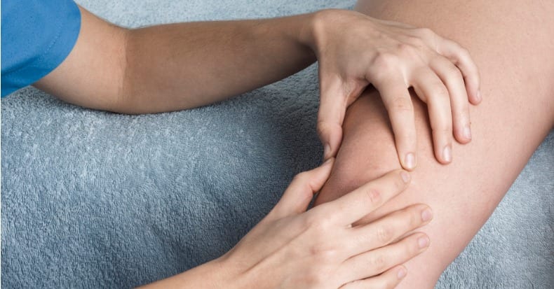
Chiropractic treatment focuses on improving function in the spine to reduce neck pain and back pain. In most cases, achieving a successful outcome is only possible when treatment addresses conditions elsewhere in the body. For example, any painful condition of the knee can change one’s gait pattern. This can result in abnormal movement in the ankle, pelvis, and lower back. It also can potentially lead to musculoskeletal pain in those areas as well. In this article, we’ll focus on patellofemoral (PF) pain, or pain that arises in the region of the knee cap. Patellofemoral pain is one of the more common knee conditions.
Anatomy of the Patella
The anatomy in and around the patella is very unique in several ways. First, the patella is the largest free-floating bone of the body. The role of sesamoid bones is to improve the function of the muscle/tendon connecting to them by optimizing the angle of action. In effect, it acts like a pulley. It significantly improves the strength and force of the muscle. The quadriceps muscles attach above the pelvis and below the upper pole of the patella. The patella glides in a grove located in the distal femur (thigh bone). A tendon then attaches the lower pole of the patella to a bony prominence located just below the knee on the shin bone.
When we flex and extend our knee, the patella slides up and down as the quadriceps contract and relax. This occurs automatically when walking, running, climbing, etc. There’s four muscles that make up the quadriceps. The rectus femoris, vastus lateralis, and vastus intermedius pull the patella up and out when we straighten the knee. Four is the vastus medialis and it pulls the kneecap up and inward. To compensate for this 3/1 disadvantage, the vastus medialis normally fires first during knee extension. This allows for proper patellar tracking and normal function.
Study on PF Pain
A 2018 study published in the Archives of Medicine and Rehabilitation looked at the “neural drive” of the four quadriceps muscles in 56 women with/without PF pain. The subjects were asked to sustain an isometric, or static knee, extension contraction at 10% of their maximum effort for 70 seconds. A specialized nerve testing tool measured the average firing rates at various time points during muscle contraction. In the non-PF pain subjects, the vastus medialis fired at higher rates. This is in comparison to the largest muscle, the vastus lateralis, which pulls the patella up and out. This was the opposite in the case of women with PF pain. Investigators suspect it may cause and/or perpetuate PF pain.
These results have led to the recommendation of isolating the vastus medialis with a specific strengthening exercise. To accomplish this, emphasize the last ten degrees of full knee extension by completely locking or straightening out the knee in extension followed by only a slight bend. Repeat 10-20 times with/without weight, depending on the degree of injury, pain, and muscle weakness. Your chiropractor can help train you in performing this exercise properly. They can also offer other highly effective exercises and treatments for knee pain.

